Learning and protecting itself: how the brain adapts
Göttingen researchers investigate the effect of certain enzymes in the healthy and diseased brain

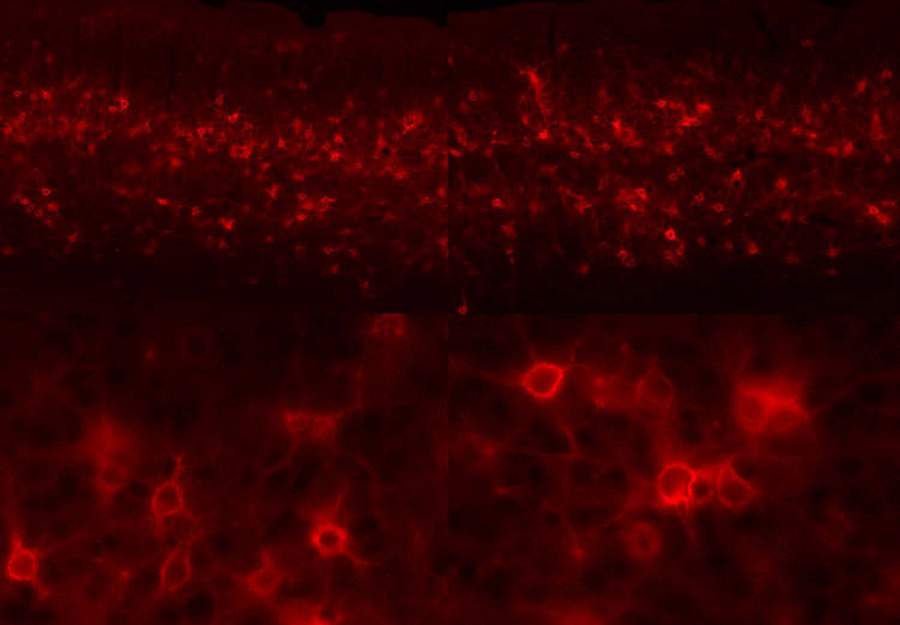
Neuronal plasticity is promoted by enzymatic degradation of the brain's extracellular matrix. This fluorescence micrograph shows neurons from the visual cortex of a mouse enveloped by red-labeled ECM molecules.
Bernstein member involved: Siegrid Löwel
/Gö, pug/ The brain is a remarkably complex and adaptable organ. However, adaptability decreases with age: as new connections between nerve cells in the brain form less easily, the brain’s plasticity decreases. If there is an injury to the central nervous system such as after a stroke, the brain needs to compensate for this by reorganising itself. To do this, a dense network of molecules between the nerve cells – known as the extracellular matrix – must loosen. This is the job of a wide variety of enzymes that ultimately regulate how plastic or how stable the brain is. Researchers at the University of Göttingen studied what happens when certain enzymes are blocked in mice. Depending on whether the brain is healthy or diseased, the inhibition had opposite effects. The results were published in the Journal of Neuroscience.
Learning and recovery from injuries depend on the plasticity of neuronal connections. Important for this are the macromolecules of the extracellular matrix, which are located between the nerve cells. When we grow up, the “stability” of this extracellular matrix increases, providing a scaffold for stabilising existing connections between the nerve cells, and consolidating what we have learned. If we experience something new, the extracellular matrix must be loosened to allow new connections to form. This relationship between stability and plasticity in the brain is regulated in the matrix with the help of enzymes such as matrix metalloproteinases (MMPs), which can ”digest” the extracellular matrix and thus “loosen” it. A team from the University of Göttingen has now been able to show in a new study that blocking the matrix metalloproteinases MMP2 and MMP9 can have opposing effects depending on whether the brain is sick or healthy.
To measure neuronal plasticity, the scientists let adult mice see only through one eye for several days and recorded the resulting activity changes in the animals’ visual cortex. In a first experiment, they examined the adaptability of the visual cortex of healthy mice in which the enzymes MMP2 and MMP9 were blocked (with SB3CT). As a result, neuronal plasticity was also blocked. In a second experiment, the team researched mice immediately after a stroke. It was already known that a stroke leads to a strong short-term increase in MMPs. In this case, the targeted, short-term inhibition of the enzymes MMP2 and MMP9 produced the opposite effect: the plasticity that had been greatly reduced by the stroke was restored, so blocking the enzymes MMP2 and MMP9 had a clear therapeutic effect.
“What made the design of our study different from many previous studies, is that the ‘matrix-degrading’ enzymes were blocked only after the experimental stroke, which simulates treatment,” says Professor Siegrid Löwel from the Department of Systems Neuroscience at Göttingen University. “We also show that the MMPs in the brain have to be very well monitored and precisely adjusted. Too low a level in the healthy brain prevents neuronal plasticity and too high a level – as after a stroke – also blocks neuronal plasticity.”
The study is part of the DFG Collaborative Research Centre 889 “Cellular Mechanisms of Sensory Processing”.
Original publication: Ipek Akol, Evgenia Kalogeraki, Justyna Pielecka-Fortuna, Merle Fricke and Siegrid Löwel. MMP2 and MMP9 Activity is Crucial for Adult Visual Cortex Plasticity in Healthy and Stroke-affected Mice. The Journal of Neuroscience (2022). 42(1): 16-32 published ahead of print: 11 November 2021, JN-RM-0902-21; DOI: https://doi.org/10.1523/JNEUROSCI.0902-21.2021


