Functional MRI for mice and humans: more direct translation of learning processes
Researchers have succeeded in identifying a network in the brain of mice that plays an important role in learning expectations and is remarkably similar to the network in the human brain.

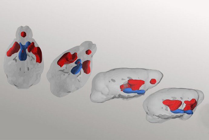
Double expectations in the brain: The figure shows the functional activation of brain regions involved in the updating of expectations from different perspectives in mice in MRI. Photo: © L. Winkelmeier und W. Kelsch
Bernstein member involved: Eleonora Russo
Functional magnetic resonance imaging (fMRI) is a standard procedure in humans to study brain networks without direct intervention in the body. It can identify active areas of the human brain depending on what the patient is doing during the examination. Among other things, fMRI can help answer neurological and psychiatric questions. This includes, for example, the role of certain networks in the brain that are important for human behaviour and learning.
However, for ethical reasons, cellular and molecular mechanisms for many psychiatric questions can only be studied in animals. For depression, psychoses and addictive disorders, for example, the learning of expectations and their correction by reality is a central mechanism that can be visualised via fMRI. But for the most important animal model – the mouse – this procedure during learning had not been available until now. Here it is important to be able to fall back on an animal model that enables a reliable transfer of knowledge. Animal models of psychiatric diseases achieve added value if they can elucidate mechanisms conserved between humans and animals. Researchers at the Central Institute of Mental Health (CIMH) in Mannheim and the Department of Psychiatry and Psychotherapy at the University Medical Center Mainz have now succeeded in doing just that.
Conserved networks encode learned reward expectations and error signals
The researchers in Mannheim and Mainz were able to show that learning of expectations and their prediction errors in mice occurs in a branched functional network that is remarkably similar to that of humans in MRI. The research team, which has now published its results in the scientific journal “Nature Communications”, was able to identify a specific network in the mouse in the MRI, in which expectations triggered by environmental stimuli are updated by recent experiences. In this network, the researchers showed for the first time that two cellular expectation signals exist in parallel in one brain area. One encodes long-term learned expectation that is robust against short-term fluctuations. The other signals expectation readily adjusted depending on what happened last.
Linking brain functions between humans and animals for better translation in psychiatric research
This new approach allows the systematic study of neural mechanisms that generate behaviour: from behavioural modelling to functional MRI that identifies functionally relevant brain areas to the activity of individual neuronal assemblies. This represents a broadly applicable method for translational, psychiatric research as well as basic research. “Behavioural MRI makes it possible, on the one hand, to determine whether conserved network mechanisms exist between humans and animal models and, on the other hand, to filter out the relevant brain regions from the multitude of possible ones and to investigate their key mechanisms,” says the team leader Univ.-Prof. Dr. Wolfgang Kelsch, Group Leader and Managing Senior Physician at the Department of Psychiatry and Psychotherapy at the University Medical Center Mainz and Group Leader at the CIMH in Mannheim. These conserved mechanisms between humans and animals should help to decipher the causes of severe psychiatric diseases and develop new therapeutic approaches.


